Breadcrumb
- Home
- Pulmonary Hypertension
- Are You a Healthcare Professional?
Are you a healthcare professional?
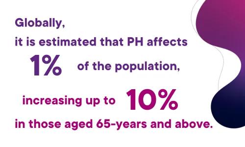
Did you know that globally it is estimated that PH affects 1% of the population, increasing up to 10% in those aged 65 years and above?2
Approximately 80% of those affected live outside of Europe and North America.2 Investigation into more accurate measures of global burden of pulmonary vascular disease is ongoing.
The most prevalent etiology worldwide of PH is due to left heart disease, particularly heart failure with preserved ejection-fraction.2,3
The leading etiology of pulmonary arterial hypertension (PAH) differs by geographic region. Idiopathic and connective-tissue associated forms tend to be the leading cause of PAH in North America and Europe.4,5,2
Sickle cell disease, schistosomiasis, and HIV infection are frequent causes of PAH in regions and countries where these diseases are endemic.6,7,2
Despite the relatively low prevalence of PH overall, more recent studies suggest that PH complicates some more common diseases, in which the development of PH is associated with increased mortality.2
| Pulmonary hypertension (PH) is a broad term encompassing many physiologic disorders that ultimately result in raised pressure in the pulmonary arteries. These disorders involve multiple clinical conditions, including cardiovascular, respiratory, and infectious diseases1. |
Definitions of PH
In order to phenotype and properly treat pulmonary hypertension, it is necessary to define hemodynamic parameters in PH. PH, in general, is defined as simply a mean pulmonary artery pressure (mPAP) of > 20 mmHg. PH is further grouped as isolated pre-capillary, isolated post-capillary, and combined types.
It is necessary to include pulmonary arterial wedge pressure (PAWP) and pulmonary vascular resistance (PVR) to help distinguish pulmonary arterial hypertension from PH due to left heart disease and other causes. Based on 2022 European Respiratory Society guidelines, pre-capillary PH is defined as a mPAP > 20 mmHg, with PAWP ≤ 15 mmHg, and a PVR > 2 Woods Units. Isolated post-capillary PH corresponds to a mPAP > 20 mmHg, with a PAWP > 15 mmHg, and a PVR < 2 WU. Combined pre- and post-capillary pulmonary hypertension is defined is a mPAP > 20 mmHg, with a PAWP > 15 mmHg, and PVR > 2 WU.1
Patients with pulmonary arterial hypertension are hemodynamically characterised by pre-capillary PH in the absence of other causes of pre-capillary PH (such as chronic thromboembolic pulmonary hypertension (CTEPH), and PH associated with lung diseases).1 It is recognised that invasive testing with right heart catheterisation is not easily accessible in many regions of the world, and there is an ongoing global discussion regarding more available means of diagnosis and the use of echocardiography.
Proceedings of the 7th World Symposium on Pulmonary Hypertension (WSPH) released an updated document in 2024 addressing the classification of PH. The thought behind such classification is to categorize clinical conditions associated with PH based on shared pathophysiologic characteristics, mechanisms, and hemodynamic parameters.1,8
- Group 1 is defined as pulmonary arterial hypertension
This is due to idiopathic, heritable, connective-tissue disease, drug/toxin exposures, HIV, portal hypertension, congenital heart disease, schistosomiasis, pulmonary veno-occlusive disease, and persistent PH of the newborn.
- Group 2 is defined as PH associated with left heart disease
This includes heart failure with reduced systolic function, preserved systolic function, cardiomyopathy, valvular heart disease, and other congenital heart conditions leading to post-capillary PH.
- Group 3 is defined as PH associated with lung disease or hypoxia
This includes chronic obstructive pulmonary disease, interstitial lung disease, combined pulmonary fibrosis and emphysema, other parenchymal lung diseases, hypoventilation hypoxia without lung disease, and developmental lung disorders.
- Group 4 is defined as PH associated with pulmonary artery obstructions
This includes chronic thromboembolic PH, and other pulmonary artery obstructions (such as from malignant and nonmalignant tumors).
- Group 5 is defined as PH with an unclear or multifactorial organisms
This often encompasses PH due to hematologic disorders (such as hemolytic anemia), systemic disorders (such as sarcoid, Langerhans’s cell histiocytosis), metabolic disorders (such as glycogen storage diseases), fibrosing mediastinitis, and pulmonary tumour thrombotic microangiopathy (PTTM).
Presenting symptoms and clinical evaluation
Elevations of pulmonary artery pressure can cause progressive increase in right ventricular (RV) afterload leading to decrease in RV contractility and eventual RV failure. The initial evaluation of patients with suspected pulmonary hypertension should begin with a detailed history and physical exam.9
PH symptoms are mainly linked to right ventricular dysfunction. The most common presenting symptom is dyspnoea on exertion. Other early signs include palpitations, fatigue, abdominal fullness, and weight gain due to fluid retention.1,9 Signs of severe or high-risk disease include syncope, haemoptysis, and cool extremities.
Historical features that may raise suspicion for PH include medical history of coronary artery disease, heart failure, chronic lung disease, connective tissue disease, or HIV.9,1
A number of social history factors are relevant including tobacco use (potentially leading to obstructive lung disease) and use of anorexigens or other toxins associated with PH (such as methamphetamine).10
A thorough family history is recommended to evaluate for familial forms of PAH, history of bleeding/clotting disorders, or recurrent miscarriage.9
Clinical evaluation beyond history and physical should include electrocardiogram and serologic testing with brain natriuretic peptide or N-terminal-pro-brain natriuretic peptide.1,11 Clinicians should consider chest radiograph to evaluate cardiac silhouette, signs of left heart disease, or evidence of parenchymal lung disease that may be contributors to PH. Pulmonary function tests (including spirometry, DLCO) and arterial blood gases may be necessary to distinguish between groups of PH.
Echocardiography can detect signs of RV pressure overload and RV dysfunction, and may help distinguish PH due to left heart disease or valvular disease.12 Ventilation/perfusion scan is recommended for the evaluation of CTEPH.13 A broader serologic and infectious evaluation may be indicated for those in whom connective tissue disease, autoimmune, hematologic, or infectious diseases are suspected to be etiologies of PH. 13
The gold standard for both diagnosing and classifying PH is with right heart catheterization. Right heart catheterization also informs which patients may be indicated for heart or lung transplant. Interpretation of right heart catheterization is recommended to be completed by a PH expert1.
As we mentioned previously, accessibility to expertise in right heart catheterization and the methodology necessary for diagnosis can be limited based on region and health care system.
This page is by no means a comprehensive summary for diagnosis and workup, but rather a snapshot of information that may be helpful for primary care providers and other healthcare workers to guide their thinking of PH in patients.
Visit ERS 2022 PH Guidelines for Diagnosis and Treatment of Pulmonary Hypertension and The Seventh World Symposium on Pulmonary Hypertension 2024 for a more detailed review. Clinicians should refer patients with suspected PH to an expert PH centre if such a referral process is available in their region.
1 - Humbert M, Kovacs G, Hoeper MM, et al. 2022 ESC/ERS Guidelines for the diagnosis and treatment of pulmonary hypertension. Eur Respir J. Published online January 1, 2022. doi:10.1183/13993003.00879-2022
2 - A global view of pulmonary hypertension - ClinicalKey. Accessed March 27, 2024. https://www-clinicalkey-com.liboff.ohsu.edu/#!/content/playContent/1-s2…;
3 - Anderson JJ, Lau EM. Pulmonary Hypertension Definition, Classification, and Epidemiology in Asia. JACC Asia. 2022;2(5):538-546. doi:10.1016/j.jacasi.2022.04.008
4 - Fling C, De Marco T, Kime NA, et al. Regional Variation in Pulmonary Arterial Hypertension in the United States: The Pulmonary Hypertension Association Registry. Ann Am Thorac Soc. 2023;20(12):1718-1725. doi:10.1513/AnnalsATS.202305-424OC
5 - Humbert M, Sitbon O, Chaouat A, et al. Pulmonary arterial hypertension in France: results from a national registry. Am J Respir Crit Care Med. 2006;173(9):1023-1030. doi:10.1164/rccm.200510-1668OC
6 - Lopes AA, Bandeira AP, Flores PC, Tavares Santana MV. Pulmonary Hypertension in Latin America: Pulmonary Vascular Disease: The Global Perspective. Chest. 2010;137(6, Supplement):78S-84S. doi:10.1378/chest.09-2960
7 - Idrees M, Butrous G, Mocumbi A, et al. Pulmonary hypertension in the developing world: Local registries, challenges, and ways to move forward. Glob Cardiol Sci Pract. 2020(1):e202014. doi:10.21542/gcsp.2020.14
8 - Kovacs, Bartolome, Denton, et al. Definition, classification and diagnosis of pulmonary hypertension. Guideline Statement | 7th World Symposium on Pulmonary Hypertension. ERS, 2024 64(4): 2104324
9 - Houston Brian A., Brittain Evan L., Tedford Ryan J. Right Ventricular Failure. N Engl J Med. 2023;388(12):1111-1125. doi:10.1056/NEJMra2207410
10 - Orcholski ME, Yuan K, Rajasingh C, et al. Drug-induced pulmonary arterial hypertension: a primer for clinicians and scientists. Am J Physiol - Lung Cell Mol Physiol. 2018;314(6):L967-L983. doi:10.1152/ajplung.00553.2017
11 - Benza RL, Gomberg-Maitland M, Elliott CG, et al. Predicting Survival in Patients With Pulmonary Arterial Hypertension: The REVEAL Risk Score Calculator 2.0 and Comparison With ESC/ERS-Based Risk Assessment Strategies. Chest. 2019;156(2):323-337. doi:10.1016/j.chest.2019.02.004
12 - Rudski LG, Lai WW, Afilalo J, et al. Guidelines for the Echocardiographic Assessment of the Right Heart in Adults: A Report from the American Society of Echocardiography: Endorsed by the European Association of Echocardiography, a registered branch of the European Society of Cardiology, and the Canadian Society of Echocardiography. J Am Soc Echocardiogr. 2010;23(7):685-713. doi:10.1016/j.echo.2010.05.010
13- Kim NH, Delcroix M, Jais X, et al. Chronic thromboembolic pulmonary hypertension. Eur Respir J. 2019;53(1). doi:10.1183/13993003.01915-2018
Find out more...

PH is a global health issue that affects all age groups.
PH is an umbrella term for elevated pulmonary artery pressures due to a wide variety of clinical disease. It is most commonly associated with left heart disease, but other forms can be idiopathic in nature, caused by congenital heart disease, connective-tissue disease, obstructive lung disease, or infection. The most prevalent etiology varies substantially by region.

Want to find out more about PVRI without becoming a member?
Are you a healthcare professional, patient or carer? Join our community as a Friend of PVRI and receive PVRi Pulse - our monthly newsletter that keeps you up to date with our latest news and developments.